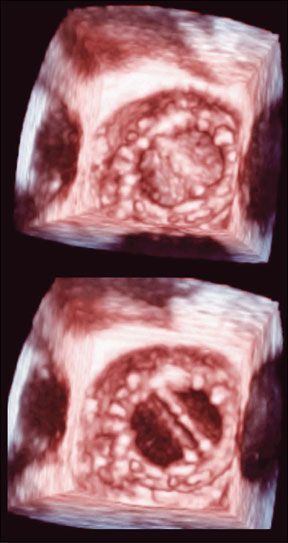
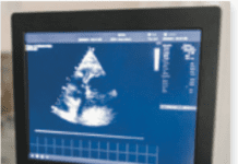
Cardiologists and cardiac surgeons are excited about a new technology that helps surgeons plan valve repair or replacement. Three-dimensional transesophageal echocardiography (3-D TEE) enables them to see the mitral valve and other heart structures in exquisite detail. "The technology provides images weve never seen before. With 3-D TEE, it is possible to better diagnose mitral valve disease or understand its pathology," says Cleveland Clinic cardiologist Takahiro Shiota, MD, an international expert on 3-D TEE and editor and author of the worlds first modern textbook on the subject of 3D echocardiography. Dr. Shiota says the new technology is so intuitive that even patients can understand it. "Most patients who see these realistic images are very impressed and express appreciation that they can better understand their mitral valve abnormalities," he says. The mitral valve is an ideal target for TEE, because it lies close to the esophagus. Patients swallow a probe tipped by a tiny ultrasound transponder, putting it within millimeters of the valve. Harmless, painless sound waves from the transponder are bounced off the valve, gathered by a computer and reconstructed into images on the screen.
To continue reading this article or issue you must be a paid subscriber.
Sign in


